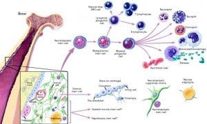
Neural Stem Cells May Replace Lost Beta Cells

The research showed how the shortage of transplantable, donated beta cells could be overcome by using stem cells to regenerate an individual’s own beta cells.

Neural Stem Cells May Replace Lost Beta CellsNeural stem cells may someday be used to replace the beta cells of the pancreas that are destroyed by diabetes, according to a Japanese research team. The findings, which were published in a recent issue of the journal “EMBO Molecular Medicine,” demonstrate how the shortage of transplantable, donated beta cells could be overcome by using stem cells to regenerate an individual’s own beta cells.

Affecting over 200 million people around the world, diabetes is caused by decreased insulin production in the pancreas—specifically, in the beta cells of the pancreas. No cure for the disease exists today, so patients must rely on supplemental insulin treatment or other therapeutics to ensure that blood glucose levels remain regulated.

The research team was headed by Dr. Tomoko Kuwabara with the AIST Institute in Tsubuka, Japan. Dr. Kuwabara’s team focused developing new ways to control stem cell differentiation in humans, which would allow scientists to transplant stem cells and give them instructions to regrow any type of cell in the human body, such as pancreatic beta cells.

Dr. Kuwabara commented that since diabetes is caused by a lack of only one type of cell, it makes a good candidate for treatment with stem cells. “As diabetes is caused by the lack of a single type of cell the condition is an ideal target for cell replacement treatments,” he said. “However donation shortages of pancreatic beta cells are a major hurdle to advancing this treatment. So a safe and easy way of using stem cells for obtaining new beta cells has been long awaited.”

Dr. Kuwabara’s team used cells from the hippocampus and olfactory bulbs—regions of the brain that scientists can easily access to obtain transplantable cells. These brain cells do not normally produce insulin in the capacity that beta cells do. However, when the neuronal cells were transplanted into diabetic rats, they began to express important characteristics normally seen in the beta cells of the pancreas. They also began to produce more insulin, resulting in a decrease of average blood glucose levels. When the transplanted cells were removed, blood glucose levels increased, revealing that the cells were responsible for the positive changes. According to the research team, this method of transplanting brain cells into the pancreas could prove to be an effective treatment for diabetes that focuses on addressing the lack of beta cells rather than medicating insulin or blood glucose levels.

“The discovery of stem cells which have virtually unlimited self-renewal raises great expectations for their use in regenerative medicine,” wrote Onur Basak and Hans Clevers, who published a close up paper in the same issue of EMBO Molecular Medicine.

“The isolation and cultivation of stem cells as a renewable source of beta cells would be a major breakthrough,” they continued.

Basak and Clevers added: “Dr Kuwabara’s team found that transplanting neural stem cells directly into the pancreas can unleash their intrinsic ability to act as critical regulators of insulin production, and most importantly they demonstrated that the cells could be gained from a patient without the need for genetic manipulation.”

“Our findings demonstrate the potential value of neural stem cells for treating diabetes without gene transfer,” said Dr. Kuwabara. “This presents an original strategy to overcome the donor shortage which has hindered cell replacement therapy.”

Dance for Health Program Encourages Activity in Youngsters

A new program is attempting to bring that message to children by encouraging children to dance away their risk of diabetes.

Dance for Health Program Encourages Activity in YoungstersPhysicians have known for decades that regular physical activity is one of the key methods of diabetes prevention, as it prevents obesity—one of the key risk factors in developing Type 2 diabetes. A new program is attempting to bring that message to children by encouraging children to dance away their risk of diabetes.

The “Dance for Health” program is put on by the University of Pennsylvania School of Nursing along with Sayre High School of Philadelphia and the Bernett Johnson Sayre Health Center. The program aims to help assess physical activity levels among school-age children and encourage them to engage in more physical activity, with the ultimate goal of reducing obesity. The program is headed by Dr. Terri Lipman, Ph.D., CRNP, of the University of Pennsylvania School of Nursing; Dr. Lipman first established the partnership between the School of Nursing and Sayre High School in 2005.

The program sees the dance team of Sayre High School leading children in an hour of dancing, once a week for a month. Researchers associated with the program use pedometers to evaluate the physical activity levels of the children, finding that they took about twice as many steps on the days that they participated in the Dance for Health program.

The children did, however, display elevated resting heart rates, even after they stopped exercise—a sign of a lack of physical fitness. Dr. Lipman hopes that the program will teach the children to engage in more frequent physical activity and improve their overall health.

“Dancing is not only free, culturally relevant, and fun, it is also an easily accessible way for children to lead a more active lifestyle,” said Dr. Lipman. “Through this program, we aim to promote to schools and health care providers the benefits of incorporating dance into children’s lives to improve their overall health.”

According to Dr. Lipman, the program’s helpful benefits have extended beyond just the children attending the dance sessions. The partnership between the School of Nursing and Sayre High School has allowed nurse practitioner students the opportunity to educate nearby communities—which are at a high risk of diabetes—on the importance of eating right, exercising frequently, and identifying warning signs in the prevention of diabetes. The partnership allows nurse practitioners to conduct basic tests such as weight, height, and waist circumference, in addition to checking for acanthosis nigricans—a condition associated with diabetes in which areas of the skin become darkened.

“Our partnership with Sayre has opened the door to a strong relationship with residents of the community around Penn,” said Dr. Lipman. “It has allowed us to work with individuals, schools, and community groups to fight diabetes together.”

The School of Nursing at the University of Pennsylvania is known as a leading research institution in the field of nursing; areas of specialty include oncology, pediatrics, geriatrics, and quality-of-life choices. The National Institutes of Health regularly grant extensive funding to the School of Nursing and many of the Master’s programs in the School of Nursing are ranked first in the U.S.

The diabetes epidemic is growing in the U.S., and shows no sign of stopping: according to the 2011 National Diabetes Fact Sheet, 25.8 million Americans have the disease, with over 18 million diagnosed cases. An astonishing 79 million people have prediabetes, a risk factor in developing Type 2 diabetes—the most common form of the disease.

TNF Inhibitors May Reduce Risk of Diabetes

According to Dr. Bili, his team’s findings support the hypothesis that TNF-a plays an important role in pathogenesis of insulin resistance—or the onset of insulin resistance, which often precedes Type 2 diabetes.

TNF Inhibitors May Reduce Risk of DiabetesA retrospective study recently published in the journal “Arthritis Care & Research” reports that tumor necrosis factor (TNF) inhibitors could help prevent diabetes in patients with rheumatoid arthritis (RA), who commonly take TNF inhibitors to help in preventing joint disease.

The study, which was funded by Amgen/Wyeth, found that adults with RA who were treated with TNF inhibitors were much less likely to develop diabetes over the three to four year period following treatment over individuals who were not treated with TNF inhibitors.

“In RA patients, use of TNF inhibitors is associated with 51% reduction in risk of developing diabetes,” said Androniki Bili M.D., M.P.H., of the Center for Health Research at the Geisinger Medical Center in Danville, Pennsylvania. Dr. Bili served as co-author on the study. He did note that retrospective studies such as this one can only determine associations between conditions and cannot positively identify causation.

The study included data from 1881 adults who had been diagnosed with RA between January 1, 2001 and December 21, 2009. Patients who already had diabetes, of which there were 294, were excluded from the study. The researchers utilized proportional hazard regression models to adjust their findings to account for a variety of confounding factors, including age, race, sex, body mass index, rheumatoid factor, and anticyclic citrullinated peptide antibodies.

After exclusions, data from 1587 patients remained. Of the cohort, 522 were users of TNF inhibitors; 91 patients had developed diabetes throughout the course of the study, among them 16 users of TNF inhibitors and 75 non-users. Diabetes incidence rates stood at 8.6 and 17.2 per 1000 person-years for TNF inhibitor users and non-users, respectively. After adjusting for covariates, researchers were left with a hazard ratio of 0.49 for users of TNF inhibitors as opposed to non-users.

“RA is a systemic inflammatory disease with major cause of death being cardiovascular disease due to accelerated atherosclerosis,” explained Dr. Bili. “Diabetes and metabolic syndrome are major risk factors for cardiovascular disease. Therefore, medications that treat both the joints and the cardiometabolic comorbidities of RA are highly desirable. Although not an official guideline, I believe that patients with RA and metabolic syndrome, insulin resistance, or [body mass index greater than] 25 kg/m2 (all risk factors for diabetes) might benefit from earlier initiation of a TNF-a inhibitor in an attempt to control the cardiometabolic comorbidities along with the joint disease.”

According to Dr. Bili, his team’s findings support the hypothesis that TNF-a plays an important role in pathogenesis of insulin resistance—or the onset of insulin resistance, which often precedes Type 2 diabetes.

The study was reviewed by Daniel Solomon, M.D., M.P.H., who had previously observed a similar decrease in diabetes risk for patients with RA who were treated with TNF inhibitors.

“The authors conducted an interesting set of analyses and came to similar conclusions as we did in a recent paper published in JAMA,” said Dr. Solomon, referring to his own similar findings. “Their data set includes some potentially important variables that our analyses could not include. Thus, this paper adds to the literature.”

However, Dr. Solomon warned that even though the studies demonstrated similar results, they did not warrant a conclusion that TNF inhibitors directly cause the reduced incidence of diabetes.

“While there is a biologic basis that may link the use of TNF inhibitors to a reduced risk of incident diabetes, 2 epidemiologic studies do not prove causation. It would be premature for doctors to incorporate these findings into their management of systemic rheumatic diseases. However, studies suggesting a link between TNF inhibitors and diabetes risk reduction speak to the inflammatory basis of diabetes and insulin resistance. This study should encourage other investigators considering immunosuppressive treatments for diabetes prevention and treatment,” said Dr. Solomon.

New Molecular Pathway Could Be Target for Diabetes Treatment

The pathway is responsible for decreases in the growth of cells that produce insulin, and is activated naturally as an individual grows older.

New Molecular Pathway Could Be Target for Diabetes TreatmentResearchers from the Stanford University School of Medicine have discovered that the artificial activation of an important molecular pathway could someday be used to treat various diseases. The pathway is responsible for decreases in the growth of cells that produce insulin, and is activated naturally as an individual grows older. The findings were published in the October 12 edition of the journal “Nature.”

The pathway being studied appears in both mice and humans. It is activated by the expression of a particular molecule called platelet-derived growth factor receptor, or PDGF-receptor for short. Expression of this molecule naturally decreases over time, as does the growth of beta cells in the pancreas, which release insulin that helps removes glucose from the bloodstream and transports it to cells where it can be used as energy.

Scientists have long known that the growth of pancreatic beta cells decreases dramatically over time. In young and newborn humans and animals, beta cell proliferation is abundant.

Another molecule known as Ezh2 appears to be involved in the pathway that reduces production of beta cells, as expression of the molecule decreases over time. Scientists were unaware, however, what caused the change in expression of Ezh2.

Researchers on the study discovered that, in juvenile mice, the expression of PDGF receptors was reduced in the islet cells of the pancreas in a similar pattern to that of the decreases in beta cell production.

Blocking the expression of PDGF receptors in young lab mice—two to three weeks old—caused decreases in Ezh2 production and the number of pancreatic beta cells than control animals whose PDGF receptors were not affected. The mice with reduced Ezh2 production also demonstrated slightly higher blood sugar levels than control subjects and could not remove glucose from the blood stream as effectively when they were given a high glycemic load, such as after a meal heavy in carbohydrates.

A lack of expression in PDGF receptors also affected fully-grown lab mice: their ability to replace beta cells was diminished when the cells were destroyed by a compound that the researchers administered to the animals. They also developed severe diabetes after their beta cells were destroyed.

“We’re hopeful that soon we might be able to manipulate this pathway in a therapeutic way in humans,” said Seung Kim, M.D., Ph.D., senior author of the study and a professor of developmental biology at Stanford University. Kim believes that scientists may one day develop a therapy that activates the pathway to promote the regrowth of beta cells or prevent their destruction.

“Perhaps by rekindling its expression and then activating it through a drug we could give in an injection or through some other route. This could be a kind of one-two punch against diabetes,” continued Kim.

If scientists can develop a therapy that promotes growth of beta cells, it could offer a new method of treating or preventing diabetes that differs from current therapeutics, which mostly focus on maintaining healthy blood glucose levels. Replacing pancreatic beta cells could allow the body to produce its own insulin to regulate blood glucose instead of relying on insulin injections or other medication to control blood sugar.

“This gives us a handhold onto a vaster problem: how to control human beta cell proliferation in a therapeutic way,”

Additionally, the researchers commented that they found other molecular patheways related to the loss of beta cells due to aging that have not yet been explored.

Analysis Suggests Metformin Reduces Risk of Colorectal Cancer

A recent meta-analysis has confirmed that patients with Type 2 diabetes who undergo metformin treatment are similarly at a significantly reduced risk of developing colorectal cancer — a 37 percent decrease in risk.

Analysis Suggests Metformin Reduces Risk of Colorectal CancerMetformin treatment has been shown in research to provide a significant decrease in risk of cancerous cell growth and thus a decrease in risk of cancer overall. A recent meta-analysis has confirmed that patients with Type 2 diabetes who undergo metformin treatment are similarly at a significantly reduced risk of developing colorectal cancer — a 37 percent decrease in risk. The results were published in the October 2011 issue of the journal “Diabetes Care,” the journal of the American Diabetes Association.

“The results indicate that metformin therapy was associated with an estimated reduction of 37% in the risk of colorectal cancer among patients with type 2 diabetes,” said the researchers on the project.

The meta-analysis was conducted at Shanghai Jiao Tong University in China and was headed by Zhi-Jian Zhang, M.D., Ph.D., with the department of epidemiology and biostatistics at the School of Public Health division of the university. Dr. Zhang’s research team pooled together the existing data from studies that investigated the effects of metformin treatment on colorectal cancer in patients with Type 2 diabetes. Information was pulled from the SciVerse Scopus and PubMed databases, comprising five studies that had been published between January 1966 and March 2011. In all, data from 108,161 patients was identified and used to analyze trends in metformin treatment as it related to risk of colorectal cancer in Type 2 diabetics.

The researchers found that metformin therapy was associated with significantly decreased risk of colorectal neoplasm when compared to patients who received non-metformin treatment (RR=0.63; 95% CI, 0.50-0.79).

The research team did not include one of the identified studies, which had been conducted on colorectal adenoma. After the exclusion of that study, the data of 107,961 diabetes patients remained, with 589 cases of colorectal cancer occurring during the follow-up periods of the four remaining studies. After the findings were adjusted to exclude the study on colorectal adenoma, researchers still found that metformin therapy was associated with significantly reduced risk of developing colorectal cancer (RR=0.63; 95% CI, 0.47-0.84).

The researchers stated that they did not find any evidence which would suggest significant heterogeneity across the studies (P=.30).

According to the research team, metformin may help fight the growth of cancer cells due to its role in the tumor suppressor pathway, where it activates AMP-activated protein kinase (9) and is also a part of the sequence that activates LKB1, a gene that is known to suppress tumors. Additionally, in vitro studies have shown that metformin actually selectively kills cancer stem cells. Those findings have been confirmed in rodent experiments, which also showed that metformin inhibited carcinoma growth in the colon.

The authors noted that previous research has tested the effects of metformin on non-diabetic humans with rectal aberrant crypt foci — one of the earliest warning signs that the individual may develop colon cancer. The findings of that study demonstrated that one month of metformin treatment was associated with a decrease in aberrant crypt foci compared to a control group who did not receive metformin treatment.

The researchers commented that if metformin therapy is definitively proven in further research to significantly reduce risk of colorectal cancer, it will likely be prescribed to most diabetics as a measure to manage blood glucose levels and also to prevent cancer.

“If metformin therapy ultimately proves effective on reducing the risk of colorectal cancer, it would likely be recommended for the overwhelming majority of diabetes patients for both blood glucose control and cancer prevention,” said the researchers.

Ten-Year Diabetics at Significantly Increased Risk of Stroke

A recent population-based study conducted by researchers at Columbia University in New York found that patients who had been diagnosed with diabetes for a period of ten years or more were over three times more likely to suffer ischemic stroke.

Ten-Year Diabetics at Significantly Increased Risk of StrokeA recent population-based study conducted by researchers at Columbia University in New York found that patients who had been diagnosed with diabetes for a period of ten years or more were over three times more likely to suffer ischemic stroke. The study, called the Northern Manhattan Study, was a large, longitudinal investigation that allowed researchers to study how risk changed over long periods of time.

Scientists have known for years that patients with diabetes are at an increased risk of ischemic stroke. However, according to Dr. Julio R. Vieira, who presented the findings at the meeting of the American Neurological Association, the ten-year length of the study allowed researchers to look at long-term risks.

The study followed 3,298 participants of varied ethnic backgrounds. The participants had no prior history of stroke. They were tested for diabetes at baseline — the beginning of the study — and then tested again every year, starting in 1993.

The mean age of the participants at the beginning of the study was 69 years, with the ages ranging from 59 to 79. Over half of the participants were Hispanic, while 24 percent were black and 21 percent were white.

At the start of the investigation, 717 patients — 22 percent — had diabetes. An additional 338 patients (10 percent) developed diabetes throughout the course of the study.

Median follow-up among the participants was nine years. Throughout the follow-up period, 244 participants were diagnosed with ischemic stroke.

Researchers used Cox proportional hazards models to determine that the participants who had diabetes at the outset of the study were 2.5 times more likely to experience ischemic stroke throughout the course of the study. Additionally, the risk of suffering an ischemic stroke increased throughout the ten-year follow up period for patients who had diabetes at baseline as well as those were diagnosed with the disease during the study. Participants who had diabetes for five years or less increased their risk of ischemic stroke to 70 percent; those who had diabetes for five to ten years had an 80 percent risk of stroke; and patients who had diabetes for ten years or more were 3.3 times more likely to have an ischemic stroke.

Dr. Vieira stated in an interview conducted after his presentation that most of the participants involved with the study had been diagnosed with Type 2 diabetes. He noted that although diabetics were at an increased risk of ischemic stroke even from the beginning of the study, that risk did not triple until the patients had diabetes for ten years. Risk of stroke was always higher in diabetics but it took a fairly long history of the disease before the risk was significantly elevated.

“Diabetes, like hypertension and all of the other risk factors for cardiovascular disease, takes a while to really cause big damage,” said Dr. Vieira. “That’s exactly what we’re seeing here.” He stressed that patients who had been newly diagnosed with diabetes still had time to make changes to reduce their risk of stroke. “You have a lot of time for intervention,” he said.

However, the risk of stroke may be what compels diabetics to begin managing the disease more effectively. Dr. Vieira commented that even though he warns diabetics of the complications that can occur as a result of the disease, including vision loss and limb amputation, it doesn’t always incur changes. Significantly increased risk of a disabling or fatal stroke, however, may be the catalyst that drives diabetics to better manage the disease.

New Study Shows Glycemic Extremes Associated with Reduced Brain Matter

Dr. Perantie’s team analyzed physiological changes in brain regions in youths who had Type 1 diabetes when they were exposed to different blood sugar levels.

New Study Shows Glycemic Extremes Associated with Reduced Brain MatterFor individuals with Type 1 diabetes, maintaining healthy blood sugar levels could be not only a necessity for long-term health, but also the key to staying mentally sharp. A study published in the online version of the journal “Diabetes” states that increased exposure to hyperglycemia — elevated levels of blood sugar — is associated with negative effects on brain size, including a decrease in whole brain gray matter. Meanwhile, hypoglycemia is associated with more severe decreases in occipital/parietal white matter volume.

The study was headed by Dana C. Perantie of the Washington University School of Medicine, located in St. Louis, Missouri. Dr. Perantie’s team analyzed physiological changes in brain regions in youths who had Type 1 diabetes when they were exposed to blood sugar levels on both the high and low ends of the spectrum. The control group for the study was comprised of the children’s siblings, who did not have Type 1 diabetes.

At baseline — the beginning of the study — each of the participants underwent brain neuroimaging. Doctors followed up with patients for two years, and patients underwent neuroimaging once again at the end of the follow-up period. Throughout the follow-up, a variety of glucose control measurements were recorded, including HbA1c levels, results from glucose meter readings, and self-reported episodes of severe hypoglycemia. After the neuroimaging was complete at the end of the follow-up, researchers studied whole brain and voxel-wise changes in the volume of gray and white matter in the study participants. Results were also adjusted for age, gender, and the age at which the patients were diagnosed with diabetes.

The findings showed no differences between patients with Type 1 diabetes and the control group in whole brain and voxel-wise brain volume throughout the two-year follow up. However, the patients in the group with Type 1 diabetes who had more hyperglycemia showed a significant decrease in whole gray brain matter than those who had less hyperglycemia. In addition, patients with Type 1 diabetes who experienced severe hypoglycemia showed significant decreases occipital/parietal white matter volume over both the control group and the group of Type 1 diabetics who had not experienced severe hypoglycemia.

“Within diabetes, exposure to hyperglycemia and severe hypoglycemia may result in subtle deviation from normal developmental trajectories of the brain,” commented the authors of the study.

The subject of changes in brain physiology related to Type 1 diabetes has been previously studied in previous research. A 2007 study published in the journal “Diabetes Care” used magnetic resonance imaging (MRIs) to study differences in brain volume among children with Type 1 diabetes and their non-diabetic siblings, age 7 to 17. The study similarly found that volume of grey and white brain matter did not differ significantly between diabetics and non-diabetics, but patients who had a history of severe hypoglycemia displayed reduced gray matter volume in the left superior temporal region. Additionally, patients who had a history of hyperglycemia showed smaller white matter volume in the right posterior parietal region and larger gray matter volume in the right prefrontal region.

The authors concluded that “qualitatively different relationships were found between hypo- and hyperglycemia and regional brain volumes in youth with type 1 diabetes.” They also commented that future research would be necessary to determine what effect, if any, the changes in brain volume would have on cognitive function and whether further exposure to extreme blood sugar conditions would have additional effects on brain matter volume.

Utah Obesity Study Shows Significant Long-Term Benefits Associated with Gastric Bypass Surgery

Patients who received surgery appeared to display better cardiovascular health, with cardiac morphology measures being significantly improved just six months after surgery.

Significant Long-Term Benefits Associated with Gastric Bypass SurgeryThe Utah Obesity Study has provided the first prospective, long-term controlled trial on the effects of gastric bypass surgery. The trial discovered that the cardiometabolic improvements associated with recent gastric bypass surgery also persist over long periods of time.

Of the 1,156 morbidly obese participants the Utah Obesity Study, 418 underwent gastric bypass surgery. An additional 417 patients sought to undergo the bariatric procedure but were unable to do so, primarily because of insufficient health insurance. Another 321 patients from the Utah Health Family Tree program were selected to function as community controls.

Doctors followed-up with patients in the Utah Obesity Study for six years. At the end of the follow-up period, patients who had undergone gastric bypass surgery maintained significant total weight loss as well as significant improvements in metabolic and cardiovascular factors compared to the control group, who did not undergo the surgery.

Physical examinations were conducted of all the patients at baseline — the start of the study — then at two years and again at six years. Exams included a wide variety of tests, including interviews with a physician and detailed medical history; submaximal exercise treadmill test and electrocardiogram; resting electrocardiograms and echocardiograms; pulmonary function; limited polysomnography; resting and exercise blood pressure; anthropometry; resting metabolic rate; urinalysis; comprehensive blood tests; and questionnaires analyzing the diet, exercise, and quality of life of the study participants. According to Dr. Ted D. Adams of the University of Utah in Salt Lake City, the six-year follow up with patients was “excellent,” with a 97% rate.

“In the surgical group, nearly all of the clinical measures improved significantly between the baseline and 2-year exams, and they remained significantly improved, compared with baseline at 6 years,” said Dr. Adams. The control groups made far less improvement than those who underwent the surgery, however: “The clinical variables in the combined control groups changed minimally if at all over the 6-year period,” continued Dr. Adams.

The group undergoing surgery showed significant weight loss that persisted throughout the six-year follow up, displaying a total weight reduction of 35 percent at two years out and 28 percent at six years out. In contrast, Dr. Adams noted, the control group displayed minimal weight loss.

The surgery group also fared better in diabetes remission at 75 percent at the six year mark, while the combined control groups were at 1 percent. At the end of the six-year follow-up, incidence of diabetes was at 2 percent for the surgery group and 16 percent for the non-surgery group.

Patients who received surgery appeared to display better cardiovascular health, with cardiac morphology measures being significantly improved just six months after surgery. Echocardiography results demonstrated reduced left atrial volume and left ventricular mass, both factors that could reduce the risk of heart failure related to obesity in the long term. Meanwhile, Dr. Adams reported that the control group displayed increased left atrial volume.

Additional improved health measures displayed by the group receiving gastric bypass surgery included reductions in waist circumference, heart rate, triglycerides, insulin resistance, systolic blood pressure, and low-density lipoprotein cholesterol that persisted through the end of the follow-up period. Higher levels of high-density lipoprotein cholesterol were also reported in the surgery group.

Dr. Adams stated that the results only support previous research on the benefits of bariatric surgery in improving health conditions related to being overweight. The findings will continue to provide insights into the benefits of the surgery and their improvements in the long term.

Dr. Adams did not disclose any conflicts of interest.

Study: Raising HDL Cholesterol Reduces Risk of Stroke and Heart Disease

The researchers discovered that, for every 5mg/dL rise in HDL cholesterol, the patients reduced their risk of hospitalization due to heart disease or stroke by 4 percent.

Raising HDL Cholesterol Reduces Risk of Stroke and Heart DiseaseA study conducted by researchers at Kaiser Permanente shows that those with Type 2 diabetes would be wise to work at improving their cholesterol. According to the findings of the study, raising HDL cholesterol — or “good” cholesterol — could be associated with a reduction in the risk of stroke and heart disease in Type 2 diabetics. The study was published in the online version of the “American Journal of Cardiology.”

Researchers arrived at their findings after reviewing medical records from 30,067 patients with Type 2 diabetes. The patients had their HDL cholesterol levels measured twice over a time period from 2001 to 2006, with the measurements being conducted from six to 24 months apart. The research team analyzed the records and reviewed the data for hospitalization due to heart disease and stroke among the patients. Then they compared HDL cholesterol data to determine whether those who improved their cholesterol between the two measurements saw any difference in hospitalization rates.

The researchers discovered that, for every 5mg/dL rise in HDL cholesterol, the patients reduced their risk of hospitalization due to heart disease or stroke by 4 percent. Additionally, an increase in HDL of 6.5mg/dL or more was associated with an 11 percent decrease in risk of hospitalization. According to the researchers, this demonstrated “that the prevention of a HDL cholesterol decrease might be at least as important as increasing the HDL cholesterol level.”

About half of all people who have been diagnosed with Type 2 diabetes also have low HDL levels, which are defined as below 40mg/dL for men and below 50mg/dL for women. According to the National Cholesterol Education Program, which is associated with the National Heart, Lung, and Blood Institute, an HDL cholesterol level of under 40mg/dL is considered to be a risk factor for heart disease and stroke.

However, the researchers at Kaiser Permanante noted that studies have still not shown that increasing HDL cholesterol levels causes a direct decrease in risk of heart attack and stroke, “perhaps because safe and effective agents that substantially increase the HDL cholesterol level remain elusive.”

According to Suma Vupputuri, investigator at the Kaiser Permanente Health Research Center in Atlanta, one possible limitation of the study is that researchers did not have access to information about what the patients did to increase their HDL cholesterol levels. It is known that lifestyle changes such as regular exercise, weight loss, and avoiding tobacco can have a positive effect on HDL. However, those changes are also known to reduce the chances of cardiovascular disease; as such, it’s unclear whether the increase in HDL or the improved lifestyles are the main catalyst behind lower risk of hospitalization.

Whether it’s improved HDL cholesterol or lifestyle changes that lead to the lower risk of hospitalization, patients are benefited either way. “I think the general message, certainly, from our paper, is yes, raising HDL is going to improve your cardiovascular outcomes,” said Vupputuri.

Steven Grover, M.D., M.P.A., director of the Cardiovascular Improvement Program at McGill University, agrees that improving HDL reduces the risk of hospitalization. “There’s enough supportive evidence from clinical trials that there’s good reason to believe raising HDL will work” to reduce that risk. Grover conducted a study in 2009 that demonstrated similar benefits in raising HDL cholesterol; however, his findings showed even greater benefits.

Barley Fiber Product Improves Post-Meal Insulin and Insulin Resistance

Branded as Barliv, the fiber was shown in tests to reduce blood glucose levels by 10.2 percent with a three-gram dose.

Insulin Resistance” src=”https://www.diabeticlive.com/wp-content/uploads/2011/10/Barley-Fiber-Product-Improves-Post-Meal-Insulin-and-Insulin-Resistance-300×300.jpg” alt=”Barley Fiber Product Improves Post-Meal Insulin and Insulin Resistance” width=”300″ height=”300″ />A new study funded by Cargill has demonstrated that beverages fortified with a type of fiber produced from barley could reduce the impact of spikes in blood sugar occurring after a meal and may also increase insulin sensitivity. The findings of the study were published in the journal “Nutrition & Metabolism.”

Branded as Barliv, the fiber was shown in tests to reduce blood glucose levels by 10.2 percent with a three-gram dose. That’s an improvement over the 7.5 percent decrease in blood sugar that study participants displayed when they received a placebo beverage.

Barliv also showed promise as a long-term treatment for decreasing insulin resistance. Participants who consumed a beverage containing six grams of Barliv every day for 12 weeks showed a 19 percent decrease in insulin resistance while a placebo group showed a 42 percent increase. Insulin resistance is the inability of the body to respond to normal levels of insulin and is a common precursor to Type 2 diabetes.

“Because this trial was designed to ensure study subjects underwent protocol-directed maintenance of body weight, it is noteworthy that the metabolic findings occurred without significant weight changes in any of the study groups,” said the researchers. The study was a joint effort conducted by scientists from Louisville Metabolic & Atherosclerosis Research Center, Cargill, the University of Kentucky, ClinData Services, and Frestedt Incorporated.

The study consisted of 50 individuals classified as “generally healthy” according to the researchers. The trial was prospective, randomized, placebo-controlled, and double-blind. Participants were separated into three groups; one group received a placebo beverage to consume daily while the other two received the beverage containing either 3 or 6 grams of Barliv. Both the placebo and Barliv beverages were raspberry-flavored.

Participants receiving the 3 gram dose of Barliv showed a 10 percent reduction in glucose levels; those receiving the 6 gram dose displayed a similar reduction, but the difference was not statistically significant. The group receiving 6 grams of Barliv had an 8 percent decrease in fasting insulin — or insulin levels between meals — while the placebo group actually showed an increase in fasting insulin.

The barley beta-glucan beverage was also associated with a reduction in body fat percentage — a 3.9 percent reduction in fat concentrations in the hips, buttocks, and thighs. However, no weight loss was recorded.

“This study supports that the 6 g/d barley beta-glucan beverage consumed over 12 weeks improves insulin sensitivity among hyperglycemic individuals who have no prior diagnosis of diabetes mellitus and no change in body weight,” said the researchers. “This study suggests barley beta-glucan may slow the deterioration of insulin sensitivity for individuals at increased risk for diabetes mellitus.”

The findings of the study could prove to be a ray of light in the fight against Type 2 diabetes, especially for those who already have pre-diabetes and are at an increased risk. The World Health Organization states that over 220 million people around the world are affected every year by diabetes, while 3.4 million die every year due to diabetes or complications from the disease. Diabetes has been on the rise for decades and the WHO is predicting that the trend won’t stop, stating that deaths from diabetes will double from 2005 to 2030.

Diabetes is a tremendous strain on health care, costing the United States alone up to $174 billion per year. A sizable portion of that number goes directly to medication for diabetes — about $116 billion of it, according to the American Diabetes Association.